Rebir Close Eyes an Open Again
Abstract
Current testify suggests that volitional opening or endmost of the eyes modulates encephalon action and connectivity. Yet, how the center land influences the functional connectivity of the primary visual cortex has been poorly investigated. Using the same scanner, fMRI data from two groups of participants like in age, sex and educational level were acquired. 1 group (n = 105) performed a resting land with eyes closed, and the other grouping (n = 63) performed a resting state with optics open. Seed-based voxel-wise functional connectivity whole-encephalon analyses were performed to study differences in the connectivity of the primary visual cortex. This region showed higher connectivity with the default way and sensorimotor networks in the optics airtight grouping, but higher connectivity with the salience network in the eyes open group. All these findings were replicated using an open source shared dataset. These results suggest that opening or endmost the eyes may set encephalon functional connectivity in an interoceptive or exteroceptive country.
Introduction
Looking for someone in a crowd, driving to an unfamiliar location, or walking past a place where there could be a dangerous animal on the loose are situations where people keep their eyes wide open. By contrast, the majority of u.s. tend to close our optics when we are trying to think or remember something. These daily life situations give u.s.a. clues that, at the brain level, in that location must be some biological mechanisms that alter and accommodate when the attentional focus is externally or internally self-driven. These changes might exist appreciable in functional connectivity (FC) networks under minimal experimental manipulation, for instance, during a resting-state condition.
Chore-related fMRI prove has shown that volitional opening or closing of the eyes during non-changing external stimulation leads to 2 different encephalon activity configurations: i associated with an "interoceptive" state (with the eyes closed), characterized past activations in areas related to imagination and multisensory action; and the other associated with an "exteroceptive" state (with the eyes open), characterized by activations in areas related to attention and ocular motor activityane,2. This change in brain activeness patterns has been shown, independently of light input3 and in early-blind individuals4, dismissing the possibility that the effects would be driven past exogenous visual stimulation. Furthermore, evidence suggests that it is an instant miracle, given that it has been found even during spontaneous center blinksfive.
Despite the relevance that this phenomenon may have in FC resting state studies, researchers take used eyes open (EO) and eyes closed (EC) settings indistinctly, with no consensus about which is more suitable depending on the aim of the research. Even more alarming, data show that in January of 2016 about 18% of the almost recently published studies did not report the approach used6. Even so, the literature indicating the existence of brain differences in both activity and connectivity based on these atmospheric condition is overwhelming. Within the scope of the functional magnetic resonance imaging (fMRI) technique, differences betwixt EO and EC conditions have been shown using a diversity of methodological approaches, such equally task fMRIane,ii,seven,8, multimodal associations with EEG data9,10,xi,12,13, spectral analysis derived measuresxiv,15,16,17,18,xix,20,21,22, regional homogeneity15,17,23, analysis of signal amplitude24,25, seed-based FC15,26,27, dynamic FC28, contained component analysis29, time-to-time fluctuations in resting-state30, Gaussian Bayesian network analysis31, and network derived measures17,32,33,34,35. Surprisingly, in spite of the large number of methodologies used inside this corpus of investigation, in that location is still a very basic question that has been poorly investigated: is it possible to observe and narrate the brain FC modulation by the middle condition just by studying the whole-brain FC of the primary visual cortex (V1)?
To the best of our knowledge, no study has been carried out to specifically answer this question. To appointment, merely one study provides direct prove in this regard26; however, the aim pursued in that investigation was different from the aim of the nowadays written report, and the evidence was based on a relatively small sample. Specifically, the primary objective of that study was to depict the relationship between brain FC at balance and brain local activeness past combining fMRI and positron emission tomography techniques. In i of the analyses presented, the authors investigated differences between EC and EO conditions in the whole-brain FC of V1 by comparing two groups of xi participants each. The results showed increased FC between V1 and salience network areas and the thalamus in the EO group, compared to the EC group. The opposite contrast (EC>EO), however, did not show statistically meaning results. Other studies have shown prove of higher FC between visual areas and the superior parietal gyrus, inferior parietal gyrus, precentral gyrus, and other visual system areas during EO, compared to EC, too as higher FC between visual areas and the precentral gyrus, postcentral gyrus, superior temporal cortex, and centre temporal cortex during EC, compared to EO15,35. Furthermore, another study showed increased effective connectivity from the salience network and central executive network to V1, besides as from V1 to the dorsal attention network, during EO but not during EC31. Yet, again, none of these studies aimed to specifically investigate differences in the FC of V1 due to/related to eye land. On the ane hand, one of them aimed to study the reproducibility of resting land data and did not directly written report V1, only rather a higher-level processing expanse in the lateral occipital cortexxv. On the other manus, the other studies aimed to investigate complex network backdrop in a specific prepare of regions, where V1 was 1 of the many regions that composed the network31,35. Thus, it is worthwhile to scientifically gather more prove about the effects of eye country on the FC of the primary visual cortex. Therefore, the object of the present study was to straight investigate the differences in the FC of V1 in EO and EC conditions by using the simplest and well-nigh intuitive resting-state FC approach: seed-based, whole-encephalon, voxel-wise FC analysis.
Taking into account the existing literature, the principal hypotheses of the present study are: 1) EC and EO conditions will modulate the FC of V1; ii) V1 volition evidence positive correlations with areas/networks typically engaged during externally driven tasks, such every bit the anterior insula and dorsal anterior cingulate (i.eastward., salience network areas), during EO; three) V1 will evidence positive correlations with areas/networks associated with introspective states (i.e. precuneus, inferior parietal cortex, medial frontal cortex) and somatosensory processing (i.due east. postcentral gyrus) during EC. To our knowledge, no study has pursued the straightforward objective of exploring the FC of V1 (independently divers and with a whole-brain voxel-wise method) in EC/EO weather condition, but its simplicity could shed low-cal on the biological grounds for the internal and external states of the encephalon.
Methods
Participants
A dataset consisting of 198 individuals (95 women; age: mean = 22.2, SD = 4.3, range = 18-40) was collected from various projects of our research group performed with the same scanner. Participants were recruited by placing posters in public places and past word of mouth. Almost of them were undergraduate students considering our research group is integrated in the campus of Universitat Jaume I of Castellón city (Spain). Three individuals were excluded due to invalid conquering, and 27 due to excessive caput motility. The final sample for the analyses included 168 participants (83 women; historic period: mean=22.01, SD=4.1, range=18-forty). Of them, 63 participants performed a resting-state session in the EO condition (EO-group), and 105 participants completed the resting-state session in the EC condition (EC-group). There were no significant differences in age (t=one.75; p=0.08), gender (χ2=0.13; p=0.72), or educational activity level (χ2=iv.five; p=0.1) between the EO and EC groups (see Tabular array one for demographics). All the participants were right-handed according to the Edinburgh Handedness Inventory36. None of them had suffered from whatever neurological or psychiatric disorders, and they had no history of head injury with loss of consciousness. Written informed consent was obtained from all participants, post-obit a protocol approved by the Institutional Review Board of Universitat Jaume I. All the study procedures conformed to the Code of Ethics of the Globe Medical Association.
Prototype acquisition
Browse sessions consisted of a resting state condition. For the EC sessions, participants were instructed to simply rest with their optics closed and not slumber or think about annihilation in detail. The same instructions, merely with the specification of keeping their optics open, were provided in the EO sessions. Just later scanning, participants were explicitly asked if they had followed the instructions and whether they had experienced any issues during the browse. None of the participants reported problems, and they all confirmed that they had followed the instructions. MRI acquisition parameters were the same as those reported in previous studies where we used part of this dataset37,38. Images were caused on a 1.5 T scanner (Siemens Avanto; Erlangen, Germany). Participants were placed in a supine position in the MRI scanner, and their heads were immobilized with cushions to reduce motion. For the resting state fMRI, a total of 200 volumes were recorded using a gradient-echo T2*-weighted echo-planar imaging sequence (TR, 2000 ms; TE, 48 ms; matrix, 64 ×64; voxel size, three.5 ×three.5 mm; flip angle, 90°; slice thickness, 4 mm; slice gap, 0.eight mm). We acquired 24 interleaved axial slices parallel to the anterior–posterior commissure airplane covering the entire encephalon. Prior to the resting country fMRI sequences, structural images were acquired using a high-resolution T1-weighted MPRAGE sequence (TR/TE = 2200/3.79 ms, flip bending 15°, voxel size = i × ane × 1 mm), which facilitated the localization and co-registration of functional information.
Epitome preprocessing
We used the Data Processing & Analysis for Brain Imaging (DAPBI V4.2, http://rfmri.org/dpabi)39, to carry out the resting state fMRI data processing. The preprocessing was similar to what was reported in one of our previous studies using this dataset37, and information technology included the post-obit steps: (ane) the first five volumes of each raw dataset were discarded to permit for T1 equilibration; (2) slice timing correction for interleaved acquisitions (the middle slice was used as the reference point); (3) head motility correction using a half-dozen-parameter (rigid body) linear transformation with a two-pass process (registered to the first paradigm and then registered to the mean of the images afterwards the first realignment); (4) co-registration of the individual structural images (T1-weighted MPRAGE) to the mean functional image; (5) Division of structural images into grey thing, white affair, and cerebrospinal fluid using the Diffeomorphic Anatomical Registration Through Exponentiated Lie algebra (DARTEL) tooltwoscore; (half dozen) removal of spurious variance through linear regression: 24 parameters from the head motion correction (6 head movement parameters, six caput move parameters 1 fourth dimension point before, and the 12 corresponding squared items)41, scrubbing within regression (fasten regression too as 1 back and 2 forward neighbors)42 at framewise displacement of (FD)>0.2 mm43, linear and quadratic trends, the white matter signal, and the cerebrospinal fluid signal; 7) spatial normalization to the Montreal Neurological Found (MNI) space (voxel size 3 × 3 × 3 mm); 8) spatial smoothing with a 4 mm FWHM Gaussian Kernel; and 8) ring-pass temporal filtering (0.01-0.1 Hz) to reduce the effect of low frequency drift and high frequency noise44,45. Given the electric current debate almost the benefits and disadvantages of including global signal regression during preprocessing of FC analyses46, we replicated all the analyses, including global signal regression in pace half-dozen described above: "Removal of spurious variance through linear regression". Furthermore, nosotros also replicated our analyses using the aCompCor method to eliminate physiological noise47,48. Specifically, we regressed out the first five components associated with white matter and cerebrospinal fluid signals. The results of these analyses are reported in the supplementary information.
Participants with more than than 1.v mm or ane.five degree of motility in any of the half dozen directions or less than 120 volumes with FD<0.2 mm (ensuring at least four minutes of rest with low FD) were excluded from the analyses. In average, participants in the terminal sample showed a mean RMS of 0.12 (SD=0.06, range=0.05-0.seven) and a mean FD of 0.14 (SD=0.04, range=0.06-0.25). No significant differences were found between the head motility metrics of the EO and EC groups.
Functional connectivity analysis
A seed-based correlation approach was used to investigate FC differences between the EO and EC groups. FC estimated with this method relies on the correlation between the boilerplate Assuming signal of a region of interest (ROI), also called a seed, and the BOLD signals of other parts of the encephalon (voxels or other ROIs). For this study, the seed region used was the V1 mask from the HCP-MMP1.0 atlas49 projected on MNI space (https://doi.org/x.6084/m9.figshare.3501911.v5). This mask was defined in a sample of 210 healthy young adults using a multimodal approach that combined data on the cortical architecture, function, connectivity, and topography (see Supplementary Fig. ane). After ROI definition, a subject-level voxel-wise FC analysis arroyo was used: for each participant, the correlation was calculated between the time series of the V1 seed and each of the time series of all the voxels of the brain. After the estimation of private correlation maps, Fisher's r-to-z transformation was performed to normalize correlation values. In society to dismiss the possibility that our results were specific to the selected atlas, we replicated our analyses using different V1 masks. Specifically, we used: (1) the Brodmann Expanse 17 obtained from the Wake Woods University PickAtlas toolbox (https://www.nitrc.org/projects/wfu_pickatlas/), which is based on the Talairach Daemon database50; (2) Brodmann Area 17 obtained from the Beefcake toolbox's maximum probability maps51, which is based on cytoarchitectonic information; (3) the maximum probability maps of V1 extracted from the ref. 52, which offers a visual cortex parcellation based on retinotopic mapping52; and (iv) calcarine mask from the Automated Anatomical Labeling (AAL) atlas53, which is based on anatomical parcellation of brain sulci. For those parcellations with more than than 1 label for V1 (east.g. left and right), we merged all the V1 masks to make a unmarried bilateral ROI. The results of this analysis are reported in supplementary material.
Grouping analyses were performed using SPM12 and Matlab R2015b (Mathworks, Inc., Natick, MA, USA). In order to determine the encephalon regions showing differential FC with V1 seed as a function of resting type, whole-encephalon, voxel-wise, 2-sample t-tests were performed. Historic period, sex, and hateful FD were included as covariates of no interest. The statistical significance threshold was prepare at p <0.05, FWE-corrected at the cluster level using a voxel-level primary threshold of p <0.001 uncorrected. Given that the results of these analyses may be driven by positive correlations, negative correlations, or a combination of the two, we performed mail service-hoc tests to further explore our results. The post-hoc tests consisted of voxel-wise, one-sample, t-exam comparisons for each group separately, within a mask of the clusters derived from the betwixt-group results. The objective of these post-hoc analyses was merely descriptive, therefore, we set up a liberal threshold of p <0.05 uncorrected.
Replication sample
We used a shared dataset acquired from the open source website http://fcon_1000.projects.nitrc.org/fcpClassic/FcpTable.html (Beijing: Eyes Open Eyes Closed Report), provided by the '1000 Functional Connectomes' Project, to replicate the results of our study. The "Beijing: optics open and optics airtight" datasetfifteen consists of 48 good for you controls (24 women; age: mean = 22.5, SD = 2.ii, range = 18–30) from a community (pupil) sample from Beijing Normal University in China. Each participant performed iii resting land fMRI scans. During the first scan (baseline), participants were instructed to rest with their eyes closed. The 2nd and third scans were randomized betwixt resting types (EO and EC). Tabular array 1 summarizes the MRI acquisition parameters of this dataset. Further details of sample characteristics and paradigm acquisition parameters are reported elsewherefifteen. Information preprocessing and FC estimation were equal to what was reported to a higher place, except that we used the prior probability distribution for affine registration of East-Asian brains during partition. One participant was excluded due to an incomplete number of volumes in the EO resting condition. Moreover, ane participant had an incomplete number of volumes in the EC resting status. Thus, for this participant, we used the baseline resting session equally the EC resting status. Afterwards preprocessing, 4 participants were excluded due to caput motion criteria. Thus, the final sample consisted of 43 participants (see Table ane for demographics).
The group analyses for this dataset consisted of whole-brain, voxel-wise, paired t-tests comparing the eyes open and eyes closed sessions. The statistical significance threshold was set at p <0.05, FWE-corrected at the cluster level using a voxel-level master threshold of p <0.001 uncorrected. Post-hoc ane-sample t-tests were likewise performed to further explore our results.
Results
Test sample
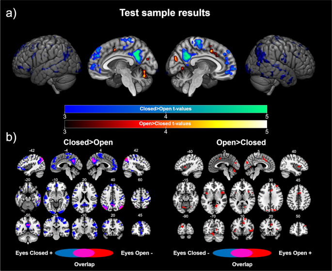
On the one mitt, the results of the analyses investigating the brain regions showing higher FC with V1 in the EC group than the EO grouping revealed statistically pregnant differences in two identifiable brain networks, namely, the default mode network, which included the bilateral junior parietal cortex, ventromedial frontal cortex, and posterior cingulate/precuneus, and the sensorimotor network, which included the bilateral postcentral and precentral gyrus. In improver, we found meaning differences in other brain regions, including the bilateral temporal cortex, bilateral junior occipital cortex, bilateral dorsolateral frontal cortex (superior, middle and junior gyrus), and dorsomedial frontal cortex (see Table two and Fig. 1a). The post-hoc comparisons showed that most all the differences were driven by positive correlations in the EC group. Interestingly, we plant a shift in the FC patterns in V1 equally a office of the resting status in the posterior regions of the default mode network (see Fig. 1b). These results indicated that V1 is correlated with the default mode network during resting with EC, but anticorrelated with this network during resting with EO.

Test sample results: (a) Differences in the functional connectivity of V1 according to eye state. Cold colors represent the encephalon regions showing higher connectivity with V1 in the eyes closed > eyes open up dissimilarity. Warm colors represent the brain regions showing higher connectivity with V1 in the optics open > eyes closed dissimilarity. The color confined stand for the t value applicative to the image. (b) Contribution of each group to the pregnant differences. Blueish colors represent the brain regions showing a correlation (positive for the contrast optics closed > eyes open, left panel; negative for the contrast optics open > eyes closed, correct panel) with V1 in the eyes closed group. Cherry colors represent the encephalon regions showing a correlation (negative for the dissimilarity eyes closed > eyes open up, left console; positive for the contrast eyes open > eyes airtight, right console) with V1 in the eyes open up group. Purple colors represent brain areas showing a shift in the direction of the correlation with V1 according to the eye state. Numbers above slices represents the corresponding MNI coordinate in millimeters.
On the other manus, when we investigated the encephalon regions showing higher FC with V1 in the EO group, compared to the EC grouping, we found significant clusters in the bilateral anterior insula and dorsal inductive cingulate cortex, which are the core regions of the salience network. Furthermore, college FC with V1 in the EO group was found in the cerebellum, bilateral superior occipital cortex, right lingual gyrus, cuneus, right heart frontal gyrus and supplementary motor area (see Table 2 and Fig. 1a). The post-hoc analyses showed that these differences were driven by positive correlations in the EO grouping.
Similar results were obtained in the analyses that included global bespeak regression and the aCompCor method during data preprocessing (see Supplementary Fig. 2). Also, the results were similar using the alternative V1 ROIs (see Supplementary Fig. 3).
Replication sample
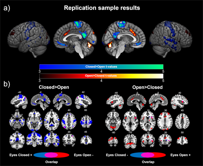
In order to validate the previously presented results, we replicated our analysis in an independent sample scanned during both EC and EO resting country conditions in the aforementioned fMRI session. Using this dataset, on the one hand, we showed significant differences in the sensorimotor network and default mode network areas in the EC> EO contrast, including the bilateral postcentral and precentral gyrus, posterior cingulate/precuneus, ventromedial prefrontal cortex, and bilateral inferior parietal cortex. We besides plant differences in the bilateral temporal cortex, bilateral inferior occipital gyrus, bilateral superior and middle frontal gyrus, and bilateral hippocampus/parahippocampus (see Tabular array 2 and Fig. 2a). The post-hoc analyses showed that the results were mainly driven by positive correlations with V1 in the EC status, with a shift in the connectivity patterns in the left inferior parietal cortex (see Fig. 2b).

Replication sample results: (a) Differences in the functional connectivity of V1 co-ordinate to heart land. Cold colors stand for the brain regions showing higher connectivity with V1 in the eyes closed > eyes open dissimilarity. Warm colors stand for the encephalon regions showing college connectivity with V1 in the eyes open> optics airtight contrast. The color bars represent the t value applicable to the paradigm. (b) Contribution of each condition to the significant differences. Blue colors represent the encephalon regions showing a correlation (positive for the contrast eyes closed > eyes open, left panel; negative for the dissimilarity eyes open > optics closed, correct panel) with V1 during the eyes closed resting state. Red colors represent the brain regions showing a correlation (negative for the contrast eyes closed > optics open up, left panel; positive for the dissimilarity eyes open > optics closed, right panel) with V1 during the eyes open resting state. Majestic colors correspond brain areas showing a shift in the management of the correlation with V1 according to the middle state. Numbers above slices represents the corresponding MNI coordinate in millimeters.
On the other hand, the results for the EO> EC contrast showed pregnant differences in the core regions of the salience network (bilateral anterior insula and dorsal anterior cingulate cortex). Furthermore, differences in the cerebellum, cuneus, bilateral superior occipital cortex, bilateral centre frontal gyrus, and thalamus were too found. The post hoc analyses revealed that the differences were driven by positive correlations in the EO condition. Evidence for a shift in the connectivity patterns was found in the correct insula.
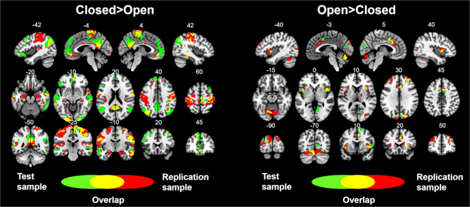
Overall, these results replicated the results observed in the examination sample (see Fig. 3). Furthermore, similar results were obtained in the analyses that included global signal regression and the aCompCor method during data preprocessing (see Supplementary Fig. 2), as well every bit in the analyses using the alternative V1 ROIs (see Supplementary Fig. three).

Overlay showing the statistically pregnant results from the test and replication samples. Green colors show the brain areas revealing pregnant connectivity with V1 in the test sample. Red colors show the brain areas revealing significant connectivity with V1 in the replication sample. Yellowish colors show the brain areas revealing significant connectivity with V1 in both the exam and replication samples. Numbers above slices represents the corresponding MNI coordinate in millimeters.
Word
The present resting-state fMRI study aimed to investigate the influence that dissimilar eye states might have on brain connectivity. The FC of the primary visual area was of item interest. To carry out this investigation, we used a test dataset consisting of 168 participants and a replication dataset consisting of 43 participants. According to our initial hypotheses, and after performing a whole-brain, voxel-wise, resting-state FC analysis, two chief results were institute: first, different eye states attune the connectivity of V1; 2nd, and more importantly, the V1's FC correlation patterns resemble a "brain's external country" and a "encephalon's internal state" during EO and EC, respectively. Overall, beyond the two datasets we found, for the resting-country EO condition, positive correlations between V1 and brain areas typically called the salience network (i.east. bilateral inductive insula and dorsal anterior cingulate cortex)54, too as the superior occipital gyrus, the cuneus, the cerebellum and the right heart frontal gyrus. For the resting-state EC condition, positive correlations were mainly plant between V1 and brain areas typically called the default style network (i.east. Posterior cingulate/Precuneus, bilateral inferior parietal cortex, and ventromedial prefrontal cortex)55 and the sensorimotor network (postcentral and precentral gyrus). Furthermore, positive correlations betwixt V1 and the bilateral temporal cortex, bilateral junior occipital cortex, and bilateral superior and middle frontal gyrus were also found during EC (encounter Fig. iii and Table two).
Our results are consistent with previous investigations showing that the different eye weather condition attune FC during resting-land acquisitions15,26,31,35. The closest and most comparable enquiry to the present report is the one conducted by Riedl et al. in 2014. In this study, the authors showed that EO increased the local action in V1, the secondary visual cortex, and the salience network regions more than EC, as evidenced by whole-brain analysis of positron emission tomography information. In parallel, resting state fMRI demonstrated an increased FC between these regions past combining visual areas and salience network areas into ii seeds. As we have replicated hither, the authors institute that during EO, the main visual area is more than coupled with salience network areas than during EC. Even so, we aggrandize these results in our study by showing that EC, compared to EO, yielded increased FC betwixt V1 and the default mode network and the sensorimotor network.
The salience network54 has been functionally related to the processing of salient inputs54,56,57. Indeed, it has been proposed that the integration of the external sensory information and the internal torso signals, including emotional information, is carried out by the salience network54,57. Some authors have plant that the right dorsal anterior insular cortex, a component of the salience network, is responsible for generating control signals that influence the activeness of the central executive network and the default mode network57,58. This same region is functionally engaged when processing high-enervating attending and control tasks, including evaluation of error, past exerting inhibition or task switching59. Thus, the salience network, and especially the dorsal anterior insular cortex, has been suggested as a encephalon switch from an introspective or internal state (due east.g., default mode network) to a readiness to reply state or external state (e.g., salience and key executive network)57.
The default style network has more often than not been associated with self-referential processing, moral data processing, and autobiographical and episodic memory retrievalthreescore,61,62,63. Other introspective or internal functions have been related to the activity of this network, such as the mind-wandering state and future self-imagination60. The default way network has been specifically investigated in the context of eye state. A previous report showed increased FC and amplitude of low frequency fluctuation (ALFF) in the precuneus and medial prefrontal cortex during EO, compared to EC18. Moreover, other studies that combined EEG and fMRI information to investigate differences between EC and EO take implicated areas of the default manner network. 1 study found negative correlations between alertness, measured equally the ratio of power in the blastoff band over the power in the delta and theta bands, and the action of the posterior cingulate gyrus, temporal gyrus, and medial frontal gyrus during resting state in the EC condition, compared to the EO condition10. Past contrast, some other study plant positive correlations betwixt the alpha power and the default mode network areas in the EO condition13. Notably, the report by Falahpour et al.10 besides showed higher positive correlations between alertness and the activity in the thalamus and insula in the EC condition when compared to the EO condition. Taking into business relationship that blastoff power is primarily recorded in occipital regions, these results are consistent with the differential design in the connectivity of V1 during EC and EO shown in our written report.
Together, the connectivity patterns shown in our written report concur with the proposal of two brain configurations associated with EO and EC, respectively: an exteroceptive state associated with attention and vigilance and an interoceptive state associated with mental imagery1,2. Thus, the fact that the main visual cortex was highly coupled with the salience network seems to indicate that, in the EO condition, the brain was in an "external fashion" or preparing to respond to an expected visual stimulus. Moreover, the connectivity pattern of V1 during the cancellation of visual input in the EC condition may involve a encephalon configuration that facilitates introspection and mental simulation, processes that have been related to activation in the default mode network and primary sensory cortex62,64.
In our opinion, the implications of these results are of import for establishing experimental fMRI assay. On the one hand, performing resting-state fMRI acquisitions in EO conditions establishes by default that at least primary visual areas volition be more than synchronized with salience network areas. On the other manus, resting-state fMRI acquisitions in EC take, by default, more synchronized connectivity betwixt visual networks and the default mode network, among others. These differences in functional synchronization may be relevant non only for resting state studies, just as well to set the control status in fMRI task studies, given the data indicating that local activity is closely related to FC in EC and EO conditions26. In this regard, our results, combined with the results of previous studies investigating the modulation of ALFF at rest based on eye state, may provide further evidence for this human relationship, given that increased ALFF in the somatosensory cortex has been consistently reported during ECfourteen,15,nineteen,20,21,22.
Finally, it should be noted that we found some differences in the results from the test and replication samples. Overall, the test sample showed greater differences in default fashion areas (larger clusters and more meaning peaks), and the replication sample showed greater differences in sensorimotor regions in the EC > EO contrast. In this same contrast, the test sample showed specific differences in inferior frontal areas and the dorsomedial frontal cortex, whereas the replication sample showed specific differences in the hippocampus and parahippocampus. Moreover, in the EO > EC contrast, the exam sample showed specific differences in the supplementary motor area and right lingual gyrus, whereas the replication sample showed specific differences in the left center frontal cortex and thalamus. Given the many differences between the 2 samples (one recruited in Spain and the other in China), it is not possible to establish the origin of these differences. Any differences could stem from methodological factors (dissimilar design, scanner, and acquisition parameters), environmental factors (dissimilar cultures), genetic factors (dissimilar ethnicity), or a combination of them. Therefore, researchers should be cautious when interpreting sample-specific results of this study in the context of the eye state.
In summary, in this written report, using two independent datasets, we accept consistently shown that the FC of V1 is modulated by the resting-state eye status. Thus, V1 showed positive FC with the salience network during the EO condition. Otherwise, V1 was positively coupled with the default manner network and sensorimotor network during EC. These results suggest that opening and endmost the eyes leads to exteroceptive and interoceptive encephalon configurations.
Information availability
The data that support the findings of this study are available from the respective author upon reasonable asking.
References
-
Marx, E. et al. Middle closure in darkness animates sensory systems. Neuroimage nineteen, 924–34 (2003).
-
Marx, E. et al. Eyes open and eyes closed as rest atmospheric condition: bear on on brain activation patterns. Neuroimage 21, 1818–24 (2004).
-
Jao, T. et al. Volitional eyes opening perturbs brain dynamics and functional connectivity regardless of light input. Neuroimage 69, 21–34 (2013).
-
Hüfner, K. et al. Differential effects of eyes open or closed in darkness on encephalon activation patterns in bullheaded subjects. Neurosci. Lett. 466, xxx–4 (2009).
-
Nakano, T., Kato, Thousand., Morito, Y., Itoi, S. & Kitazawa, Due south. Blink-related momentary activation of the default way network while viewing videos. Proc. Natl. Acad. Sci. U. S. A. 110, 702–six (2013).
-
Waheed, S. H. et al. Reporting of Resting-Land Functional Magnetic Resonance Imaging Preprocessing Methodologies. Brain Connect six, 663–668 (2016).
-
Qin, P. et al. Cocky-specific stimuli interact differently than non-self-specific stimuli with eyes-open versus eyes-closed spontaneous activeness in auditory cortex. Front. Hum. Neurosci vii, 437 (2013).
-
Wiesmann, M. et al. Eye closure in darkness animates olfactory and gustatory cortical areas. Neuroimage 32, 293–300 (2006).
-
Ben-Simon, E., Podlipsky, I., Arieli, A., Zhdanov, A. & Hendler, T. Never resting brain: simultaneous representation of two alpha related processes in humans. PLoS One 3, e3984 (2008).
-
Falahpour, Thousand., Chang, C., Wong, C. Due west. & Liu, T. T. Template-based prediction of vigilance fluctuations in resting-country fMRI. Neuroimage 174, 317–327 (2018).
-
Feige, B. et al. Cortical and subcortical correlates of electroencephalographic alpha rhythm modulation. J. Neurophysiol. 93, 2864–72 (2005).
-
Henning, S., Merboldt, K.-D. & Frahm, J. Task- and EEG-correlated analyses of BOLD MRI responses to optics opening and closing. Brain Res. 1073–1074, 359–64 (2006).
-
Mo, J., Liu, Y., Huang, H. & Ding, Thousand. Coupling between visual alpha oscillations and default mode activeness. Neuroimage 68, 112–8 (2013).
-
Liang, B. et al. Encephalon spontaneous fluctuations in sensorimotor regions were directly related to eyes open and eyes airtight: evidences from a automobile learning approach. Front end. Hum. Neurosci eight, 645 (2014).
-
Liu, D., Dong, Z., Zuo, X., Wang, J. & Zang, Y. Eyes-open/eyes-closed dataset sharing for reproducibility evaluation of resting state fMRI data assay methods. Neuroinformatics 11, 469–76 (2013).
-
McAvoy, Grand. et al. Resting states affect spontaneous Assuming oscillations in sensory and paralimbic cortex. J. Neurophysiol. 100, 922–31 (2008).
-
Wei, J. et al. Eyes-Open and Eyes-Closed Resting States With Opposite Encephalon Activeness in Sensorimotor and Occipital Regions: Multidimensional Evidences From Auto Learning Perspective. Forepart. Hum. Neurosci 12, 422 (2018).
-
Yan, C. et al. Spontaneous brain activity in the default mode network is sensitive to different resting-state weather condition with limited cerebral load. PLoS One 4, e5743 (2009).
-
Yang, H. et al. Amplitude of low frequency fluctuation inside visual areas revealed past resting-state functional MRI. Neuroimage 36, 144–52 (2007).
-
Yuan, B.-K., Wang, J., Zang, Y.-F. & Liu, D.-Q. Amplitude differences in high-frequency fMRI signals between optics open and eyes closed resting states. Front. Hum. Neurosci eight, 503 (2014).
-
Zhou, Z., Wang, J.-B., Zang, Y.-F. & Pan, G. PAIR Comparison betwixt Two Within-Grouping Conditions of Resting-State fMRI Improves Classification Accuracy. Front end. Neurosci eleven, 740 (2018).
-
Zou, Q. et al. Detecting static and dynamic differences between eyes-closed and eyes-open resting states using ASL and BOLD fMRI. PLoS One 10, e0121757 (2015).
-
Vocal, Ten. et al. Frequency-Dependent Modulation of Regional Synchrony in the Human Encephalon by Eyes Open and Eyes Closed Resting-States. PLoS One x, e0141507 (2015).
-
Bianciardi, M. et al. Modulation of spontaneous fMRI activity in homo visual cortex past behavioral state. Neuroimage 45, 160–8 (2009).
-
Wong, C. W., DeYoung, P. Northward. & Liu, T. T. Differences in the resting-state fMRI global signal amplitude betwixt the eyes open and eyes closed states are related to changes in EEG vigilance. Neuroimage 124, 24–31 (2016).
-
Riedl, V. et al. Local activeness determines functional connectivity in the resting homo brain: a simultaneous FDG-PET/fMRI written report. J. Neurosci. 34, 6260–6 (2014).
-
Zou, Q. et al. Functional connectivity between the thalamus and visual cortex under eyes closed and eyes open atmospheric condition: a resting-state fMRI study. Hum. Brain Mapp. xxx, 3066–78 (2009).
-
Wang, 10.-H., Li, Fifty., Xu, T. & Ding, Z. Investigating the Temporal Patterns within and betwixt Intrinsic Connectivity Networks under Optics-Open and Eyes-Airtight Resting States: A Dynamical Functional Connectivity Study Based on Phase Synchronization. PLoS Ane 10, e0140300 (2015).
-
Agcaoglu, O., Wilson, T. W., Wang, Y.-P., Stephen, J. & Calhoun, Five. D. Resting state connectivity differences in eyes open versus eyes closed weather condition. Hum. Brain Mapp. 1–11, https://doi.org/x.1002/hbm.24539 (2019).
-
Li, Z., Zang, Y.-F., Ding, J. & Wang, Z. Assessing the mean strength and variations of the fourth dimension-to-time fluctuations of resting-land brain activity. Med. Biol. Eng. Comput. 55, 631–640 (2017).
-
Zhang, D. et al. Directionality of large-scale resting-state brain networks during eyes open and eyes airtight conditions. Front. Hum. Neurosci ix, 81 (2015).
-
Li, Z. et al. Effects of resting state condition on reliability, trait specificity, and network connectivity of brain part measured with arterial spin labeled perfusion MRI. Neuroimage 173, 165–175 (2018).
-
Patriat, R. et al. The effect of resting condition on resting-state fMRI reliability and consistency: a comparing between resting with eyes open up, closed, and fixated. Neuroimage 78, 463–73 (2013).
-
Van Dijk, K. R. A. et al. Intrinsic Functional Connectivity As a Tool For Human Connectomics: Theory, Properties, and Optimization. J. Neurophysiol. 103, 297–321 (2010).
-
Xu, P. et al. Different topological organisation of homo brain functional networks with eyes open up versus eyes closed. Neuroimage 90, 246–55 (2014).
-
Oldfield, R. C. The assessment and analysis of handedness: the Edinburgh inventory. Neuropsychologia 9, 97–113 (1971).
-
Adrián-Ventura, J., Costumero, V., Parcet, M. A. & Ávila, C. Reward network connectivity 'at rest' is associated with reward sensitivity in healthy adults: A resting-state fMRI study. Cogn. Affect. Behav. Neurosci. 19, 726–736 (2019).
-
Adrián-Ventura, J., Costumero, V., Parcet, One thousand. A. & Ávila, C. Linking personality and encephalon anatomy: a structural MRI approach to Reinforcement Sensitivity Theory. Soc. Cogn. Affect. Neurosci fourteen, 329–338 (2019).
-
Yan, C.-G. G., Wang, Ten.-D., Di, Zuo, X.-N. North. & Zang, Y.-F. F. DPABI: Data Processing & Analysis for (Resting-State) Encephalon Imaging. Neuroinformatics fourteen, 339–51 (2016).
-
Ashburner, J. A fast diffeomorphic image registration algorithm. Neuroimage 38, 95–113 (2007).
-
Friston, Chiliad. J. K. J., Williams, S., Howard, R., Frackowiak, R. S. J. R. S. & Turner, R. Motility-related furnishings in fMRI time-series. Magn. Reson. Med. 35, 346–55 (1996).
-
Yan, C.-G. et al. A comprehensive cess of regional variation in the impact of caput micromovements on functional connectomics. Neuroimage 76, 183–201 (2013).
-
Power, J. D., Barnes, Grand. A., Snyder, A. Z., Schlaggar, B. Fifty. & Petersen, S. E. Spurious only systematic correlations in functional connectivity MRI networks arise from subject move. Neuroimage 59, 2142–54 (2012).
-
Biswal, B., Yetkin, F. Z. Z., Haughton, Five. Yard. V. Thou. & Hyde, J. Due south. J. S. Functional connectivity in the motor cortex of resting human brain using repeat-planar MRI. Magn. Reson. Med. 34, 537–41 (1995).
-
Lowe, M. J., Mock, B. J. & Sorenson, J. A. Functional connectivity in single and multislice echoplanar imaging using resting country fluctuations. Neuroimage 7, 119–132 (1998).
-
Tater, K. & Flim-flam, 1000. D. Towards a consensus regarding global betoken regression for resting country functional connectivity MRI. Neuroimage 154, 169–173 (2017).
-
Behzadi, Y., Restom, K., Liau, J. & Liu, T. T. A component based noise correction method (CompCor) for BOLD and perfusion based fMRI. Neuroimage 37, 90–101 (2007).
-
Muschelli, J. et al. Reduction of motion-related artifacts in resting state fMRI using aCompCor. Neuroimage 96, 22–35 (2014).
-
Glasser, One thousand. F. et al. A multi-modal parcellation of human cerebral cortex. Nature 536, 171–178 (2016).
-
Lancaster, J. Fifty. et al. Automated Talairach atlas labels for functional encephalon mapping. Hum. Brain Mapp. ten, 120–131 (2000).
-
Eickhoff, Due south. B. et al. A new SPM toolbox for combining probabilistic cytoarchitectonic maps and functional imaging data. Neuroimage 25, 1325–35 (2005).
-
Wang, L., Mruczek, R. E. B., Arcaro, M. J. & Kastner, S. Probabilistic maps of visual topography in homo cortex. Cereb. Cortex 25, 3911–3931 (2015).
-
Tzourio-Mazoyer, North. et al. Automated anatomical labeling of activations in SPM using a macroscopic anatomical parcellation of the MNI MRI single-subject encephalon. Neuroimage 15, 273–89 (2002).
-
Seeley, W. W. et al. Dissociable intrinsic connectivity networks for salience processing and executive command. J. Neurosci. 27, 2349–56 (2007).
-
Raichle, M. Due east. et al. A default mode of brain function. Proc. Natl. Acad. Sci. USA 98, 676–82 (2001).
-
Kurth, F., Zilles, K., Fox, P. T., Laird, A. R. & Eickhoff, Due south. B. A link between the systems: functional differentiation and integration within the man insula revealed by meta-analysis. Brain Struct. Funct. 214, 519–534 (2010).
-
Uddin, L. Q. Salience processing and insular cortical function and dysfunction. Nat. Rev. Neurosci. sixteen, 55–61 (2015).
-
Sridharan, D., Levitin, D. J. & Menon, V. A critical role for the right fronto-insular cortex in switching between central-executive and default-way networks. Proc. Natl. Acad. Sci. Us 105, 12569–74 (2008).
-
Chang, L. J., Yarkoni, T., Khaw, K. W. & Sanfey, A. One thousand. Decoding the role of the insula in man cognition: functional parcellation and large-calibration reverse inference. Cereb. Cortex 23, 739–49 (2013).
-
Raichle, M. Eastward. The restless encephalon: how intrinsic activity organizes brain function. Philos. Trans. R. Soc. Lond. B. Biol. Sci 370, 20140172 (2015).
-
Andrews-Hanna, J. R. The brain'south default network and its adaptive role in internal mentation. Neuroscientist 18, 251–lxx (2012).
-
Andrews-Hanna, J. R., Smallwood, J. & Spreng, R. North. The default network and self-generated idea: component processes, dynamic control, and clinical relevance. Ann. N. Y. Acad. Sci 1316, 29–52 (2014).
-
Buckner, R. Fifty., Andrews-Hanna, J. R. & Schacter, D. L. The encephalon'due south default network: anatomy, function, and relevance to disease. Ann. N. Y. Acad. Sci. 1124, 1–38 (2008).
-
Kosslyn, Southward. M., Ganis, G. & Thompson, W. Fifty. Neural foundations of imagery. Nat. Rev. Neurosci. ii, 635–42 (2001).
Acknowledgements
This work was supported by grants from Generalitat Valenciana (AICO/2018/038) and Ministerio de Economía y Competitividad (PSI2016–78805-R) to CA. Additionally, this work was supported past a pre-doctoral graduate program grant (FPU15/00825) to JA; a post-doctoral graduate program grant (APOSTD-2018 from the Generalitat Valenciana and the European Social Fund: "Investing in your time to come") to EB; and a post-doctoral graduate program grant (FJCI-2014-19589) to VC. Financial back up for the replication sample data used in this project was provided by a grant from the National Natural Science Foundation of Red china (30770594) and a grant from the National High Engineering science Plan of China (863: 2008AA02Z405). The authors declare no competing interests.
Writer information
Affiliations
Contributions
5.C., C.A. and J.A.V. conceived and designed the written report. Five.C., Due east.B. and J.A.V. performed the experiment. Five.C. and J.A.Five. analyzed the data. 5.C., C.A. and E.B. wrote the newspaper. All authors revised the commodity and gave concluding approval of the version submitted.
Respective writer
Ethics declarations
Competing interests
The authors declare no competing interests.
Additional information
Publisher's note Springer Nature remains neutral with regard to jurisdictional claims in published maps and institutional affiliations.
Supplementary information
Rights and permissions
Open Access This article is licensed under a Artistic Eatables Attribution iv.0 International License, which permits use, sharing, adaptation, distribution and reproduction in any medium or format, as long as y'all requite appropriate credit to the original author(s) and the source, provide a link to the Creative Commons license, and betoken if changes were fabricated. The images or other 3rd party cloth in this commodity are included in the article'due south Creative Commons license, unless indicated otherwise in a credit line to the fabric. If textile is not included in the commodity'due south Artistic Commons license and your intended use is not permitted past statutory regulation or exceeds the permitted employ, you will need to obtain permission directly from the copyright holder. To view a re-create of this license, visit http://creativecommons.org/licenses/past/4.0/.
Reprints and Permissions
About this article
Cite this article
Costumero, Five., Bueichekú, East., Adrián-Ventura, J. et al. Opening or closing eyes at rest modulates the functional connectivity of V1 with default and salience networks. Sci Rep x, 9137 (2020). https://doi.org/10.1038/s41598-020-66100-y
-
Received:
-
Accepted:
-
Published:
-
DOI : https://doi.org/10.1038/s41598-020-66100-y
Further reading
Comments
Past submitting a comment you agree to bide past our Terms and Community Guidelines. If you find something abusive or that does not comply with our terms or guidelines please flag it as inappropriate.
Source: https://www.nature.com/articles/s41598-020-66100-y
0 Response to "Rebir Close Eyes an Open Again"
Post a Comment